Adenoidectomy Pictures: Before and After Recovery Images
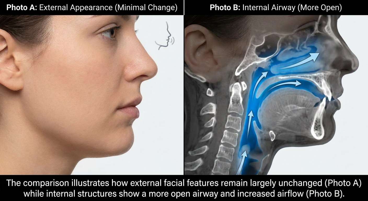
It’s understandable to search for adenoidectomy pictures before a child’s surgery (or after it) to reduce uncertainty: What do enlarged adenoids look like? What does “removed” look like? And how can you tell if healing is on track? This guide walks through the most common image types you’ll see online—“before” views of obstruction, intraoperative endoscopic images, and “after” healing appearances—plus a realistic recovery timeline and what symptoms are often expected in the first week. One quick reassurance: many meaningful “after” changes happen internally in the back of the nose (the nasopharynx). So, while families often look for before-and-after face photos, external pictures usually do not show major visible changes after adenoid surgery. These symptoms are often described in patient guidance and may occur during recovery. [2]
If you’d like to read more about the symptoms that bring families to evaluation in the first place, see our related guide: signs of enlarged adenoids in kids https://sleepandsinuscenters.com/blog/big-adenoids-symptoms-in-kids-key-signs-every-parent-should-know.
Bottom line: internal changes matter most after adenoidectomy, and external photos rarely tell the full story.
What Are Adenoids (and Where Are They)?
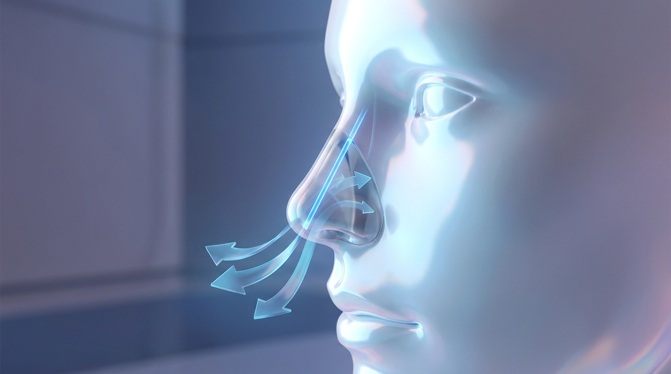
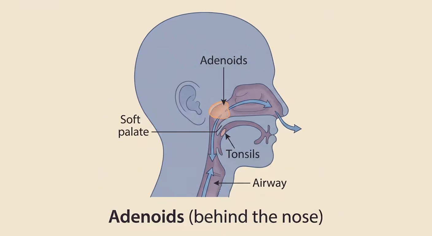
Adenoids are a patch of lymphoid tissue located behind the nose, high in the throat, in an area called the nasopharynx. They are not usually visible by simply looking in the mouth (unlike tonsils).
A helpful analogy: think of the adenoids as a soft “pad” of immune tissue sitting above the back of the throat—close enough to affect airflow, but tucked out of sight.
Because of their position, enlarged adenoids can:
- Narrow airflow behind the nose
- Contribute to snoring or mouth breathing
- Be associated with ear pressure, recurrent ear infections, or fluid behind the eardrum
Graphic idea: A side-view head diagram labeling: nasal cavity, adenoids (nasopharynx), soft palate, tonsils, and the airway.
Takeaway: adenoids live out of sight, which is why endoscopic views—not mouth photos—are most useful.
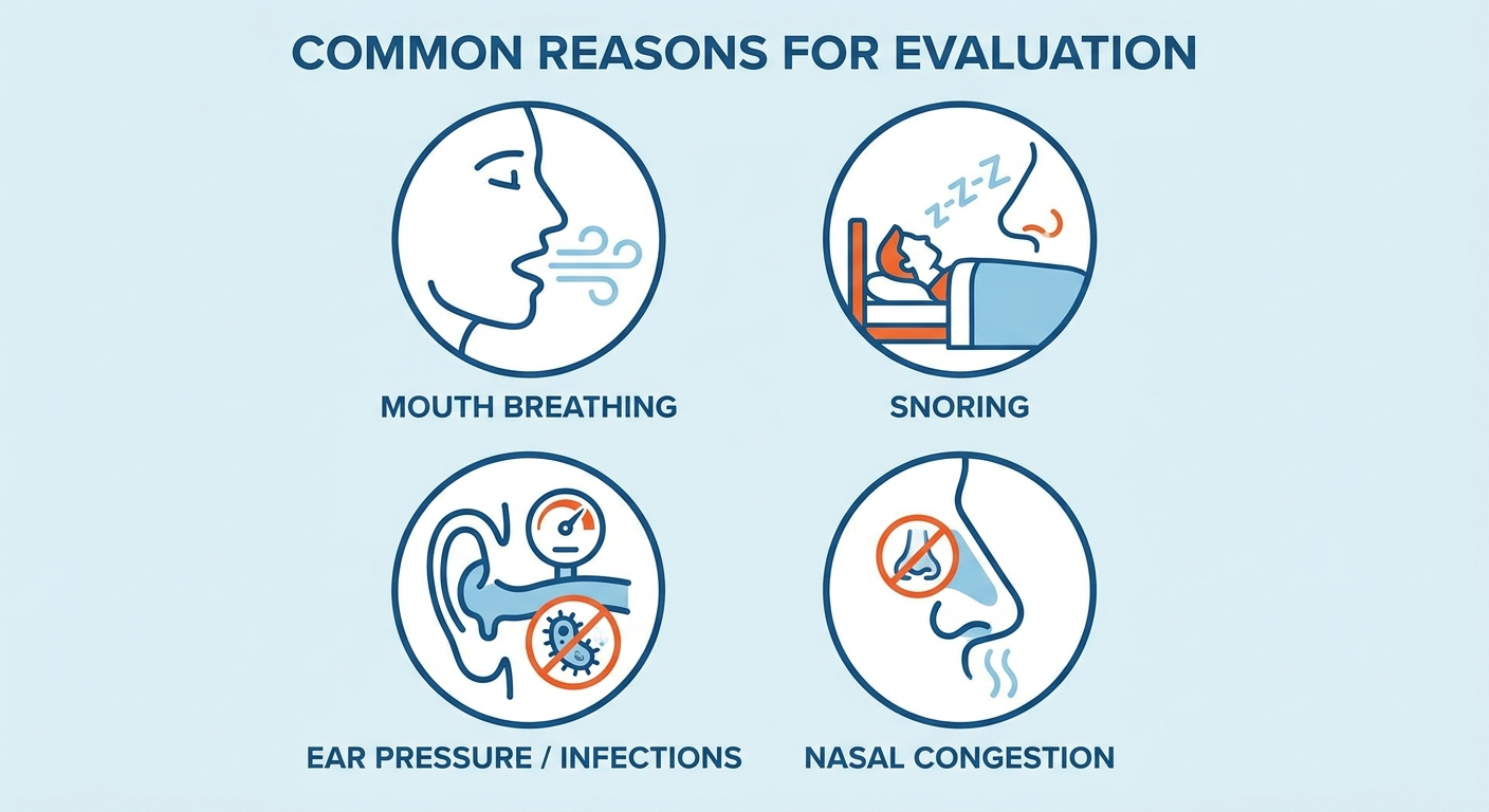
Symptoms That Often Lead to Adenoidectomy (Why the “Before” Looks the Way It Does)
When people look for adenoidectomy pictures, they’re often trying to connect symptoms to something visual. Common “before” symptom patterns include:
- Ongoing nasal congestion and mouth breathing
- Snoring or sleep-disordered breathing
- Recurrent ear infections or persistent fluid behind the eardrum
- Recurrent sinus/nasal infections
In day-to-day terms, parents often describe a child who “always sounds stuffy,” sleeps with an open mouth, or seems to breathe more comfortably sitting upright than lying flat.
For families dealing with nighttime obstruction, you may also find this helpful: can’t breathe through the nose at night https://sleepandsinuscenters.com/blog/cant-breathe-through-nose-at-night.
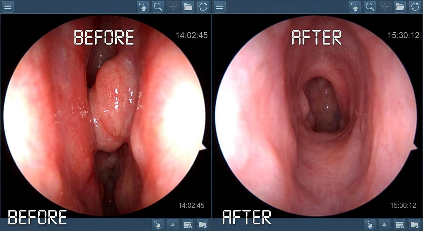
When photos are especially helpful
- A clinician shares an enlarged adenoids image from a nasal endoscopy to show the level of blockage
- You’re comparing “crowded” versus “open” space behind the nose to understand the goal of surgery
A practical example: an endoscopic “before” image may show the adenoid tissue filling much of the space behind the nose, while an “after” image shows a clearer passage for airflow.
Key point: pictures can help with understanding, but they cannot confirm whether an individual child’s healing is normal.
Types of Adenoidectomy Pictures You’ll See Online (and What Each One Means)
Online adenoid removal pictures tend to fall into three main categories. Understanding which type you’re viewing helps set expectations—especially because “before and after” often refers to endoscopic views, not facial photos.
1) “Before” pictures: enlarged adenoids
Common formats
- Educational drawings (clear and non-graphic)
- Endoscopic views (a small camera looking into the nasal passages toward the nasopharynx)
- Occasionally X-ray-style illustrations (used historically; endoscopy is more direct today)
What to notice
- Degree of obstruction: partial narrowing vs. near-complete blockage
- Inflammation and mucus: swelling can make tissue look larger than it would on a “good day,” and secretions can make images look more dramatic
If you’re comparing images online, keep in mind that lighting, camera angle, and mucus can make two similarly sized adenoids look very different. These “before” visuals are often what people mean when they search for adenoidectomy pictures—they want to see what “enlarged” looks like compared with normal airflow space.
2) Intraoperative pictures: endoscopic adenoidectomy views
Many modern sets of endoscopic adenoidectomy photos are taken during surgery using an endoscope for visualization. These images may show:
- The adenoid tissue (“adenoid pad”)
- Surgical instruments
- Energy-assisted devices (depending on technique)
- The surgical field as tissue is reduced/removed
Expectation-setting: intraoperative images can look intense because they’re clinical close-ups. Their purpose is education and documentation, not aesthetics. Open-access surgical articles sometimes include high-resolution figures that clearly show technique and the surgical view. [1]
3) “After” pictures: the postoperative nasopharynx
When you see adenoidectomy before and after comparisons using endoscopy, the “after” view typically shows the nasopharynx after adenoidectomy with:
- A noticeably more open airway behind the nose
- Mild crusting or superficial cautery changes early on
- Healing tissue that can look uneven at first
A key point: “after” does not always mean “perfectly smooth.” Early healing can look patchy or crusted before it settles—similar to how a scab looks “messy” before it resolves.
Summary: the most meaningful “after” photo usually shows simply more clear, open space for airflow.
True “Before and After” Expectations (What Changes—And What Doesn’t)
Changes you can expect
In many cases, the “after” difference is best described as functional and internal:
- A clearer space for nasal breathing on endoscopy
- Less mouth breathing and snoring in many children (depending on the cause)
- Fewer infections in selected patients (results vary by underlying factors)
If you’re looking at picture sets, the most meaningful “win” is often simply more visible open space behind the nose.
Changes you usually won’t see in external photos
Even if internal airflow improves significantly, external “before and after” face photos often show little to no visible difference:
- No meaningful cosmetic change is expected from adenoid surgery alone
- The main goal is improving airflow and related ENT concerns—not changing appearance
Bottom line: adenoidectomy is about function, not facial appearance.
Modern Techniques Featured in Recent Image Sets (Why Newer Photos Look Different)
Endoscopic adenoidectomy (common in recent series)
Endoscopic approaches can produce clearer “before/after” pictures because visualization of the nasopharynx is direct, and hard-to-see areas can be assessed more thoroughly. This is one reason newer image sets may look more “zoomed in” and detailed than older illustrations.
Energy-assisted approaches (example: radiofrequency)
Some newer surgical series illustrate energy-assisted methods in very clear intraoperative images. In one recent endoscopic radiofrequency series, reported averages included:
- Mean operative time around 14–15 minutes
- Mean blood loss around ~31 mL [1]
These figures are not universal and may not apply to every patient.
Takeaway: technique influences both the surgical view and the clarity of “before/after” images.
Safety, Risks, and What Complications Might Look Like in Photos
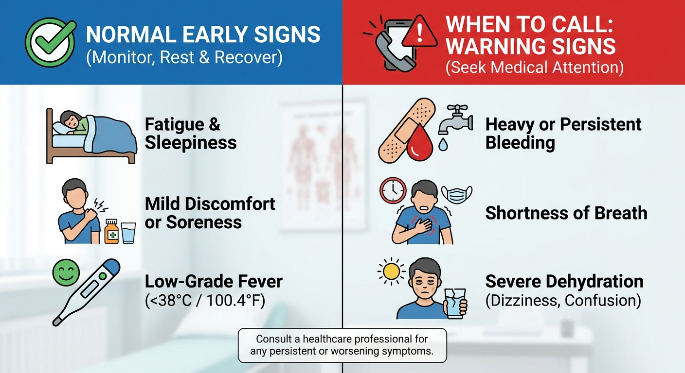
Common, normal early symptoms (not usually photographed)
Many normal early experiences are symptoms rather than photo-worthy changes, including:
- Mild throat soreness
- A “blocked nose” feeling
- Temporary bad breath (halitosis) [2]
Parents sometimes worry that bad breath means infection; however, patient resources often describe it as a short-term healing issue that improves as the surgical area recovers. [2]
Bleeding and other complications (rare, but important)
While serious issues are uncommon, online photo searches sometimes focus on “what complications look like.” In general educational terms, concerning signs discussed in post-op instructions often include:
- Bright red bleeding from the nose or mouth
- Vomiting blood
- Symptoms that worsen after initially improving
Reported complication rates (context, not a promise)
In one large endoscopic series, reported rates included:
- Primary bleeding: ~0.3%
- Secondary bleeding: ~0.1%
- Regrowth requiring revision: ~0.5% over one year [1]
These numbers provide context from one study; individual risk depends on many factors and may differ in other settings.
Reminder: post-op guidance from your surgical team should drive decision-making, not internet photos.
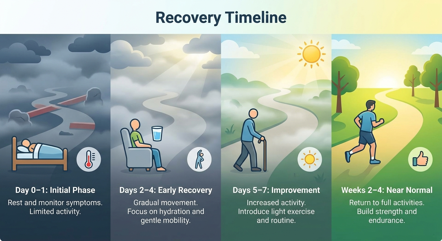
Adenoidectomy Recovery Images—What Healing May Commonly Look Like (Timeline)
When people search for adenoidectomy recovery images, they’re usually trying to match an expected timeline. Here’s what many patient resources describe, paired with what an internal endoscopic view might show in broad terms. Photos and diagrams are for education only and cannot determine whether an individual child’s healing is normal; symptoms and follow-up with the surgical team matter most.
Day 0–1 (surgery day and first night)
- Common experiences: grogginess, sore throat, nasal congestion.
- Possible endoscopic appearance: early swelling and mild surface changes where tissue was treated.
Days 2–4
- Common experiences: stuffiness, mouth breathing at night, mild ear pressure.
- Bad breath can be more noticeable during this window and then improve as healing continues. [2]
Days 5–7
- Many patients report improvement around this point, though recovery varies. Endoscopic photos (when shown in journals) may look less crusted with a more clearly open airway.
Weeks 2–4 (often pictured in journals rather than patient galleries)
- The lining often appears smoother and less irritated as residual healing changes fade.
Key point: recovery is a process; day-to-day swelling can change how the nasopharynx looks and feels.
How to Use Adenoidectomy Pictures Wisely (Avoid Misleading Comparisons)
Why your “after” may not match someone else’s
Two “after” photos can look different even when both are normal, because of:
- Age and starting adenoid size
- Allergy or infection-related inflammation at the time of surgery
- Technique differences (traditional vs. endoscopic/energy-assisted)
- Whether other procedures were performed at the same time
A simple way to think about it: pictures capture one moment in time, but recovery is a process—and swelling can vary day to day.
Red flags that should prompt a call (even if pictures online look “similar”)
General post-op instructions often emphasize contacting your surgical team if there are concerning symptoms like fever that persists, dehydration (not drinking), significant bleeding, or breathing difficulty. [2]
Bottom line: use pictures for education—not diagnosis—and call your care team for concerns.
Where to Find Reliable Adenoidectomy Pictures (High-Quality Sources)
Patient-friendly image libraries (simpler, labeled)
Look for labeled diagrams and non-graphic educational pages. Some hospital education pages also compile galleries of enlarged adenoid examples. [4]
High-resolution intraoperative figures (more technical)
For detailed endoscopic adenoidectomy photos, open-access journal articles (such as those hosted on PubMed Central) often include figure captions that explain what you’re seeing. [1]
Privacy and ethics reminder
Avoid sharing identifiable patient images publicly. If your clinic took endoscopic photos during evaluation, you can ask whether they can be reviewed at follow-up (many clinics can display them for education).
Tip: prioritize reputable sources and ask your care team to interpret images in your child’s context.
Treatment Overview (Brief, for Context)
Non-surgical management that may be tried first (case-dependent)
Depending on the situation, treatment may start with options aimed at inflammation or contributing factors, with observation when symptoms are mild.
When surgery is considered
Adenoidectomy is typically considered when symptoms persist—such as nasal obstruction/sleep concerns or recurrent ear/sinus issues—despite appropriate evaluation and initial management. [2]
If you’re also comparing procedures, you may like: adenoidectomy vs tonsillectomy https://sleepandsinuscenters.com/blog/adenoidectomy-vs-tonsillectomy-for-children-which-20260316181610.
Takeaway: the decision to operate depends on symptoms, impact on quality of life, and clinical findings.
Lifestyle & Home Tips for a Smoother Recovery (Patient-Friendly)
Recovery instructions vary by surgeon and patient, but general guidance commonly emphasizes:
- Hydration and easy-to-swallow foods in the early days
- Using pain relief only as directed by your surgical team
- Rest, then a gradual return to school and activities as permitted
- Avoiding smoke and irritants during healing
Some families find nighttime humidity helpful if recommended by their clinician; others won’t need any special nasal care after surgery.
Simple habits—fluids, rest, and following instructions—support smoother healing.
FAQs
Can I see my adenoids in my child’s mouth?
Usually not. Adenoids sit behind the nose (in the nasopharynx), so they typically aren’t visible by looking in the mouth.
Why does my child’s breath smell bad after adenoidectomy?
Temporary bad breath can be part of normal healing and is commonly mentioned in patient recovery guidance. [2]
How long does adenoidectomy recovery take?
Many patients feel significantly better in about a week, though internal healing can continue beyond that. [2]
Do adenoids grow back?
Regrowth is uncommon but possible. One recent endoscopic series reported revision for regrowth in about 0.5% of patients over one year. [1] These figures are study-specific and not universal.
Are endoscopic adenoidectomy pictures “better” than older pictures?
Endoscopic images are often clearer because they visualize the nasopharynx directly, making before-and-after airway space easier to interpret.
Book an appointment
If you’d like an ENT specialist to review symptoms, walk you through what adenoid images typically show, or discuss whether adenoidectomy is appropriate, you can book an appointment at https://www.sleepandsinuscenters.com/. Next step: a focused consultation can personalize what the images mean for your child.
Medical disclaimer
This article is for educational purposes only and is not medical advice. Please consult a qualified healthcare provider for diagnosis and treatment.
References
1. Aouf M. et al. A 7-year experience in endoscopic radiofrequency adenoidectomy. PubMed Central (2025). https://pmc.ncbi.nlm.nih.gov/articles/PMC12491561/
2. NHS. Adenoidectomy. (Last reviewed 10 March 2023). https://www.nhs.uk/tests-and-treatments/adenoidectomy/
3. Adenoidectomy (Adenoid Removal). Patient education resource. https://my.clevelandclinic.org/health/treatments/15447-adenoidectomy-adenoid-removal
4. Liv Hospital. Adenoidectomy Pictures: View Enlarged Adenoids. https://int.livhospital.com/adenoidectomy-pictures-view-enlarged-adenoids/
Don’t let allergies slow you down. Schedule a comprehensive ENT and allergy evaluation at Sleep and Sinus Centers of Georgia. We’re here to find your triggers and guide you toward lasting relief.