Nasal Polyps Images: What They Look Like and How to Identify Them
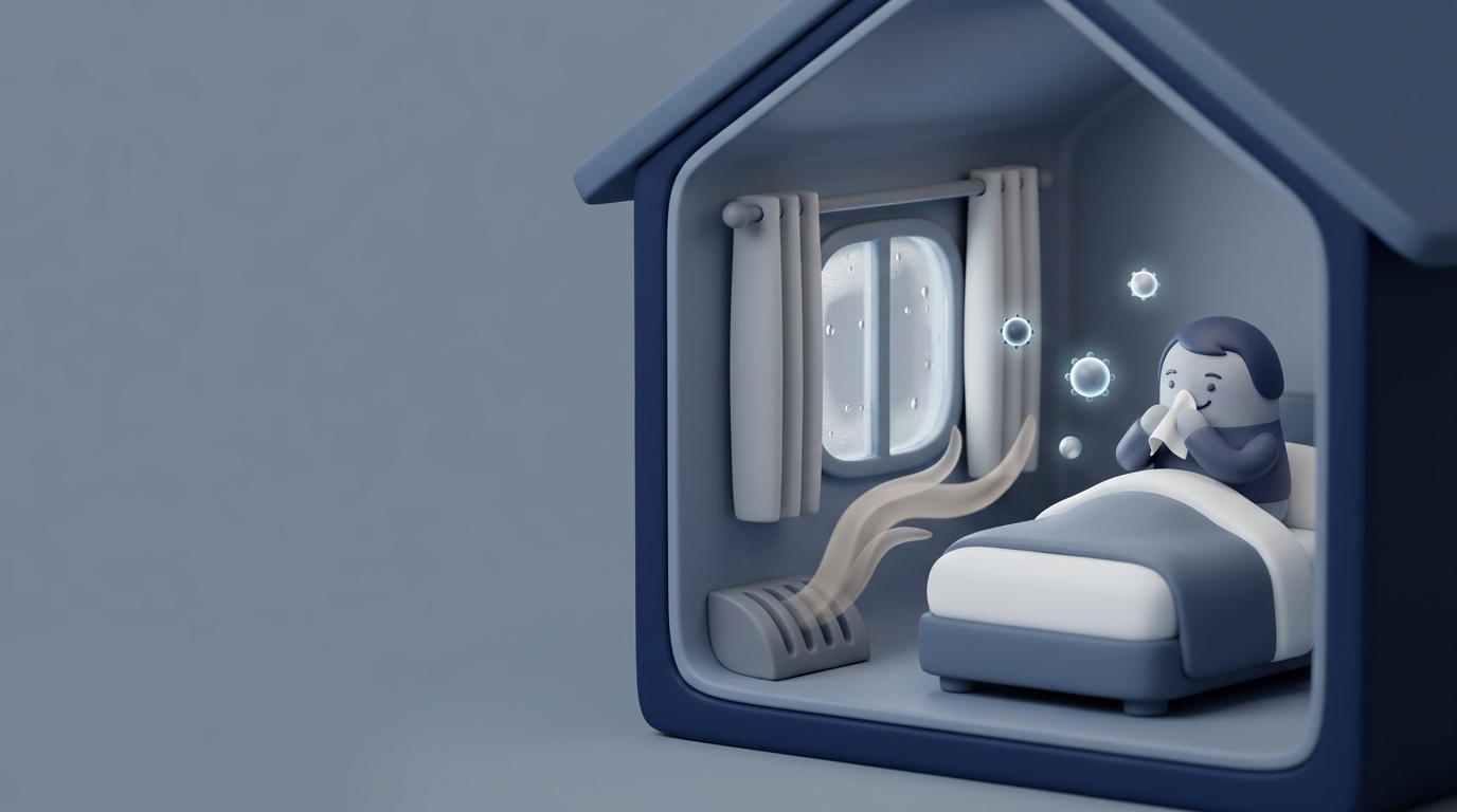
Searching for nasal polyps images is a common first step when you’re dealing with long-lasting congestion, sinus pressure, or changes in smell. Pictures can help you understand what polyps typically look like—but they can’t confirm what’s happening in your nose or sinuses.
Below, we cover what nasal polyps often look like on different exams (in-office views vs. CT imaging), common symptoms, “look-alike” conditions, and when it makes sense to see an ENT at Sleep and Sinus Centers of Georgia for an accurate diagnosis.
For a deeper explainer on basics, read: https://sleepandsinuscenters.com/blog/what-are-nasal-polyps
Quick Answer — What Do Nasal Polyps Look Like?
Typical appearance (in plain language):
- Soft and smooth (not crusty or sharp)
- Pale/gray and gel-like
- Teardrop-shaped or like grapes in a cluster
- Often painless (even if they cause pressure or blockage)
A simple way to picture them: less like a solid bump and more like a smooth, slightly translucent water balloon or peeled grape sitting in the nasal passage. This “pale, smooth, grape-like” description appears consistently in medical references.¹²
Why images can look different person to person:
- Size and number (one small polyp vs. many)
- Location (nasal cavity vs. deeper sinuses)
- How the image was taken (basic exam, endoscopy, or CT)²⁴
In short, typical images are helpful for recognition, but they’re not a perfect template to diagnose yourself.
Important Note Before You Compare Images Online
Online photos can’t confirm a diagnosis. Many other conditions can resemble polyps in a photo, such as enlarged/swollen turbinates, cysts or severe inflammation, benign growths (e.g., papillomas), and—rarely—tumors that should not be missed.²³
A common example: swollen turbinates can fill space and, in a still photo, look like a mass when they’re actually normal anatomy that’s inflamed. Lighting, angle, and camera type can also change how tissue appears, especially in endoscopy images.²³
When self-checking is unsafe: do not attempt to examine or remove nasal tissue at home, as this can cause bleeding, irritation, or infection. The safest way to get a clear answer is a professional exam.
Bottom line: use photos as a learning tool, but rely on a professional exam for diagnosis.
Nasal Polyps Images — What They Look Like in Different Exams
What you see depends on how you’re looking.
What polyps look like on a nasal exam (anterior rhinoscopy)
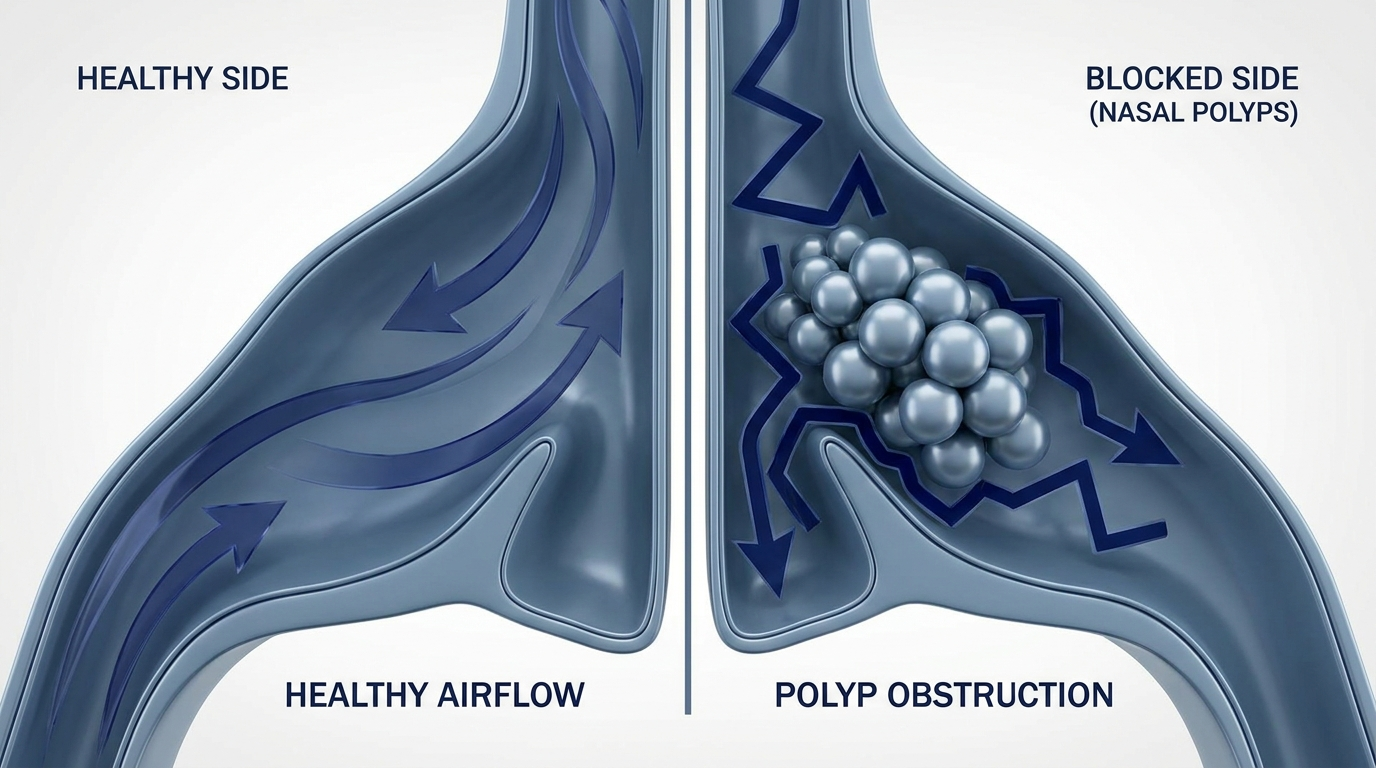
During a routine exam (looking into the nostril with a light), polyps may be visible if they’re large and positioned forward. They often appear as smooth, pale masses that don’t match the surrounding pink nasal lining.¹² Because many polyps extend deeper near sinus drainage pathways, a basic exam can miss them—especially early on.

What polyps look like on nasal endoscopy images
A nasal polyp endoscopy image is often the most recognizable picture patients see online. With a small camera (endoscope), ENTs can view deeper parts of the nasal cavity where polyps commonly appear. Typical descriptions include pale, translucent tissue; teardrop or grape-like shapes; a gelatinous look; and tissue that may appear to hang from sinus openings into the nasal passage.¹⁴ Learn more about the experience: https://sleepandsinuscenters.com/blog/what-is-nasal-endoscopy----and-is-it-painful

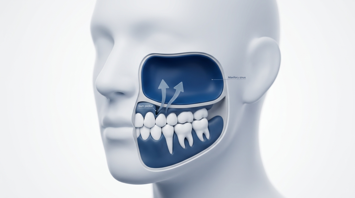
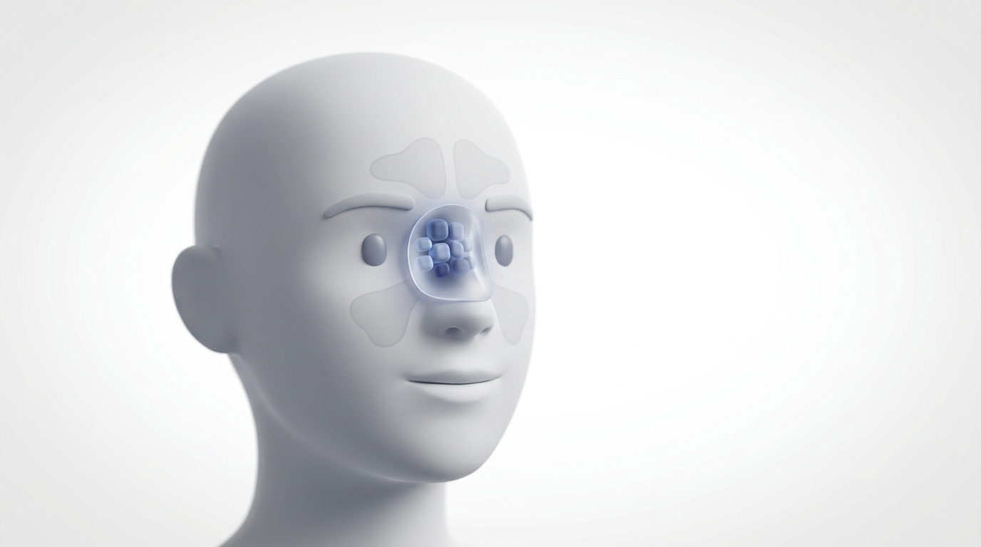
What polyps look like on a CT scan
A CT scan is useful when symptoms suggest disease deeper in the sinus cavities. On CT, nasal polyps are usually seen indirectly as soft-tissue density or sinus opacification, which may reflect polyps and/or associated inflammation and secretions.³⁴ More on why CT matters: https://sleepandsinuscenters.com/blog/how-sinus-ct-scans-help-ent-diagnosis

Additional CT findings with sinonasal polyposis
In long-standing or extensive disease, CT may also show enlarged infundibula (widened drainage pathways) and bony remodeling or thinning of adjacent sinus walls.³ These details help plan treatment safely—especially if a procedure is being considered. Think of endoscopy as showing the surface details and CT as mapping the deeper terrain.

How to Tell If It Might Be a Nasal Polyp (Symptoms Checklist)
Common symptoms linked with ongoing inflammation include nasal congestion or blockage, postnasal drip, reduced or lost sense of smell/taste, facial pressure/fullness, and runny nose.²⁴ Many describe the blockage as always there, and others notice smell loss first.
Clues it’s more than a cold: colds improve within days to a couple of weeks. With polyps, symptoms often last weeks to months, feel consistently blocked rather than alternating sides, and include persistent or worsening smell loss.² If symptoms persist beyond a typical cold, get evaluated.
What Causes Nasal Polyps?
The underlying driver is chronic inflammation in the nose and sinuses, often in the setting of chronic rhinosinusitis.²⁴ Polyps don’t appear overnight; they develop where the lining stays irritated and swollen over time.
Common associated conditions and risk factors include allergies or chronic rhinitis, asthma, possible association with aspirin-exacerbated respiratory disease (AERD), and recurrent or chronic sinus disease.²⁴ Managing the underlying inflammation is key to long-term control.

Conditions That Can Look Like Nasal Polyps in Pictures (Differential Diagnosis)
Common look-alikes include turbinate swelling, mucus retention cysts, benign tumors (e.g., papillomas), and malignancy (uncommon, but important to rule out). A deviated septum isn’t a lesion but can crowd or obscure the view and make tissue appear mass-like.
Why imaging helps: CT clarifies what’s happening inside the sinuses, whether there are changes involving bone, and provides the anatomic roadmap needed for safe surgical planning.³⁴ Because different problems can look similar in photos, endoscopy and CT often provide decisive clarity.
How ENTs Diagnose Nasal Polyps (What to Expect at the Visit)
Step 1 — Symptom history and nasal exam: review duration, smell/taste changes, and prior treatments.
Step 2 — Nasal endoscopy (in-office camera exam): closer look at polyp tissue, drainage pathways, mucus, and inflammation.¹² Often the quickest way to connect symptoms with anatomy.
Step 3 — CT scan (when needed): evaluate extent of disease and help plan treatment, including procedures when appropriate.³⁴ A focused history, endoscopy, and CT—when needed—create a complete, accurate picture.
Treatment Options (From Least to Most Involved)
Medical treatments (often first-line): steroid nasal sprays, short courses of oral steroids in select cases (clinician-guided), and saline rinses as supportive care.²⁴ Consistent daily therapy often matters more than occasional rescue treatment.
Biologic medications (for certain severe cases): for eligible patients with chronic rhinosinusitis with nasal polyps (CRSwNP), coordinated by ENT and, when appropriate, allergy/pulmonology teams.⁴
Procedural/surgical options: polyp removal and/or endoscopic sinus surgery when large, persistent, or recurring despite medical therapy; imaging guides planning and safety.²³ Learn more: https://sleepandsinuscenters.com/blog/how-ent-doctors-remove-nasal-polyps
What results to expect (and recurrence reality): many patients experience meaningful relief with the right combination of therapies, but polyps can recur. Smaller polyps may shrink with treatment; persistent polyps often need ongoing management.²⁴
Lifestyle & At-Home Tips to Support Symptom Control
Helpful daily habits (alongside medical care) may include consistent saline irrigation, allergen reduction when allergies are involved, and managing asthma/allergies to reduce overall airway inflammation.² Avoid overusing decongestant sprays and any DIY removal attempts.
When to See a Doctor (and Red Flags)
Make an ENT appointment if you have nasal blockage plus smell loss lasting more than a few weeks, symptoms that keep returning despite typical treatment, or ongoing concerns after comparing images and noticing a strong symptom match.
Seek urgent evaluation for one-sided bleeding or severe one-sided obstruction, new facial swelling, vision changes, severe headache, or neurologic symptoms. Book with Sleep and Sinus Centers of Georgia: https://www.sleepandsinuscenters.com/
FAQs
What do nasal polyps look like inside the nose? Pale, smooth, soft, grape-like or teardrop-shaped; usually best seen on endoscopy.¹²
Can you see nasal polyps without an endoscope? Sometimes, if they’re large and forward. Many are deeper and easier to identify with endoscopy and/or CT.¹³
What do nasal polyps look like on a CT scan? Usually indirect: soft-tissue density or sinus opacification reflecting polyps and/or inflammation/secretions.³
Are nasal polyps dangerous? Usually benign, but can significantly affect breathing and smell. Other conditions can look similar, so evaluation matters.²³
Can nasal polyps go away on their own? Smaller polyps may shrink with treatment; persistent polyps often need ongoing management.²⁴
Conclusion — Use Images as a Starting Point, Not a Diagnosis
Online images can help you recognize the typical look: pale, smooth, grape-like tissue on endoscopy and filled or gray sinus spaces on CT. But many conditions can mimic polyps, and some polyps hide deeper in the sinuses. Use images as a starting point for an ENT evaluation, not a diagnosis. For clarity and a personalized plan, book at https://www.sleepandsinuscenters.com/.
References
1. Ubie Health. Nasal Polyps Appearance Inside Nose — https://ubiehealth.com/doctors-note/nasal-polyps-appearance-inside-nose-view-science2753q3
2. Cleveland Clinic. Nasal Polyps — https://my.clevelandclinic.org/health/diseases/15250-nasal-polyps
3. Radiopaedia. Sinonasal polyposis — https://radiopaedia.org/articles/sinonasal-polyposis?lang=us
4. PMC. Chronic Rhinosinusitis with Nasal Polyps: Review — https://pmc.ncbi.nlm.nih.gov/articles/PMC11213496/
This article is for educational purposes only and is not medical advice. Please consult a qualified healthcare provider for diagnosis and treatment.
Don’t let allergies slow you down. Schedule a comprehensive ENT and allergy evaluation at Sleep and Sinus Centers of Georgia. We’re here to find your triggers and guide you toward lasting relief.