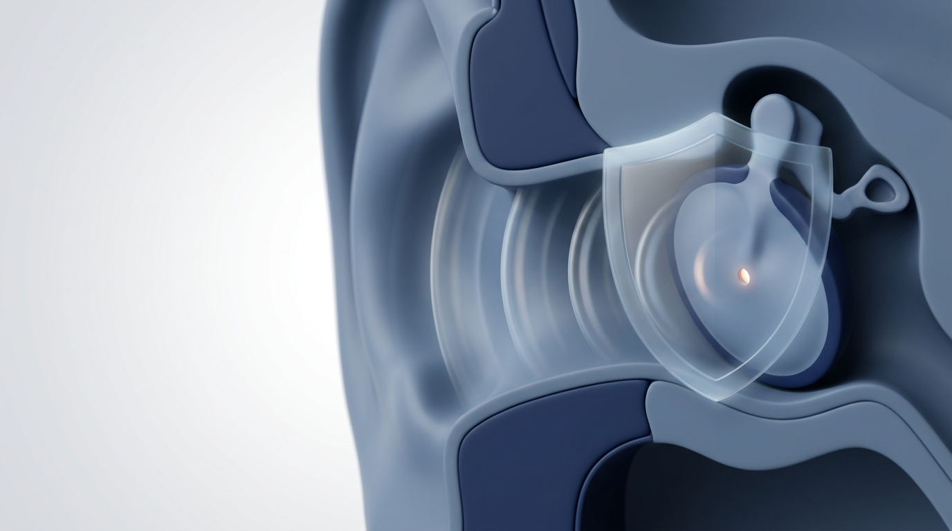

When Imaging Alone Isn’t Enough: Sinus Disease Diagnosis and Treatment Options
Introduction — Why a “Clear” Scan Doesn’t Always Mean “No Sinus Problem”
When people think about sinus disease diagnosis, they often picture a scan—usually a CT scan (and sometimes an MRI). Imaging is commonly ordered because it can show sinus anatomy, blockage patterns, and details that matter for planning procedures. CT is excellent at answering questions like, “How narrow are the drainage pathways?” or “Which sinuses look inflamed?”
But imaging doesn’t tell the whole story. A meaningful sinus disease diagnosis usually comes from putting several pieces together: your symptoms and their pattern, a focused exam, and often nasal endoscopy—and in selected situations, sinus biopsy. That combination helps avoid missed diagnoses and also helps prevent treating “scan findings” that aren’t actually causing problems. Clinical correlation is emphasized in major imaging guidance for sinonasal disease.
A simple way to think about it: a scan is a snapshot, but sinus symptoms are a movie. If the snapshot is taken on a “good week” (or right after starting medications), the image may look better than how you feel over time.
In this article, you’ll learn:
- Which symptoms should guide evaluation (and which can be misleading)
- Why CT findings can be overcalled or undercalled
- How nasal endoscopy and biopsy fill critical gaps
- “Red flag” sinus symptoms that deserve urgent evaluation
- Common sinus treatment options matched to the underlying cause
Educational note: This article is for general education and isn’t a substitute for personalized medical care.
In short: a clear or mildly abnormal scan doesn’t rule out a real problem—and a “busy” scan doesn’t always mean you need treatment.

Common Symptoms That Should Guide Sinus Disease Diagnosis (Not Just the Scan)
Typical symptoms (often overlap with allergies/colds)
Many sinus complaints overlap with viral colds and allergic rhinitis, including:
- Nasal congestion or obstruction
- Facial pressure/fullness
- Thick nasal drainage or post-nasal drip
- Reduced smell or taste
- Cough (often worse at night), fatigue, bad breath
These symptoms matter because a scan can’t tell how you feel day to day—or how symptoms fluctuate with seasons, exposures, sleep, hydration, or treatments.
Symptoms that suggest chronic vs acute patterns
The pattern is often more informative than a single “snapshot” image:
- Acute: symptoms lasting less than 4 weeks—most are viral and self-limited; antibiotics are generally not indicated unless bacterial infection is strongly suspected (for example, symptoms persist/worsen after about 7–10 days, or there’s severe onset with high fever and purulent discharge).
- Chronic sinusitis: symptoms lasting 12+ weeks, or repeated cycles of improvement and relapse.
For example, someone might report “pressure and thick drainage every spring,” while another person feels congested “pretty much all the time” with only brief improvement. Those stories can point the workup in very different directions—even if the CT images look similar.
Why symptoms alone can be misleading
- Migraine, TMJ disorders, and nerve pain can mimic “sinus headache.”
- Allergic rhinitis can cause major congestion and drainage without true sinus infection.
- Reflux can contribute to throat symptoms that feel “sinus-related.”
Bottom line: symptoms guide the process best when they’re paired with a targeted exam and, when appropriate, endoscopy and imaging.
What Causes Sinus Disease? (And Why the Cause Changes the Workup)
Inflammatory causes
A large share of chronic sinus problems are inflammation-driven, often influenced by:
- Allergic rhinitis
- Non-allergic triggers (irritants, pollution, smoke)
- Nasal polyps (chronic rhinosinusitis with nasal polyps vs without polyps)
Inflammation can narrow drainage pathways over time—sometimes without a “classic infection” picture—so the best testing strategy often focuses on confirming what’s actually happening in the nasal cavity.
Structural/anatomic contributors
Anatomy can set the stage for poor drainage and repeated inflammation, such as:
- Deviated septum
- Turbinate enlargement
- Narrow drainage pathways in the osteomeatal complex
Think of anatomy like tight plumbing—if the openings are narrow, even minor swelling can tip things into blockage.
Infectious causes
Not every flare is bacterial. Causes can include:
- Viral infections (common early on)
- Bacterial infections (selected cases)
- Fungal sinus disease (distinct categories with very different management)
Less common but important causes
Some findings require extra attention because they change the urgency and workup:
- Benign growths
- Tumors (uncommon overall, but important not to miss—especially with unilateral symptoms)
Key idea: similar symptoms can come from very different causes, so testing should be matched to the suspected driver.

The Role of Imaging (CT/MRI): What It Does Well
Why CT is often the first choice for sinus imaging
A sinus CT scan is widely used because it shows bony anatomy and patterns of sinus aeration and blockage. It’s also a cornerstone of surgical planning if procedures are being considered. For a deeper look at what the test can and can’t show, see: Sinus CT Scan: What It Shows and How It Helps Diagnose Sinus Issues — https://sleepandsinuscenters.com/blog/sinus-ct-scan-what-it-shows-and-how-it-helps-diagnose-sinus-issues
What a sinus CT scan can show
CT may reveal:
- Mucosal thickening
- Partial or complete sinus opacification
- Air-fluid levels (often more suggestive of acute infection)
- Soft tissue density that may represent polyps or inflammation (often non-specific)
- Septal deviation, turbinate hypertrophy, or narrowed drainage pathways
These findings are useful—but they don’t automatically equal “disease that needs treatment.” Interpretation should always be in clinical context.
Takeaway: CT is excellent for anatomy and patterns, but it’s only one piece of the diagnostic puzzle.
Why Imaging Alone Isn’t Enough (The Core Problem)
CT “false positives” are common
A “false positive” CT scenario happens when CT shows mucosal thickening or partial opacification, yet the person has minimal symptoms—or symptoms driven by something else. Importantly, mucosal thickening is nonspecific and can be seen during a cold, allergy season, dental issues, or even in people without symptoms.
CT can miss early or complex disease—especially in high-risk patients
Imaging can also under-detect problems in certain situations. In high-risk patients (for example, those who are immunocompromised), early invasive fungal sinusitis may have subtle findings and can be difficult to separate from other serious causes based on imaging alone. When the story doesn’t fit, a broader evaluation is needed.
Imaging can’t reliably tell what the tissue is
CT often describes “soft tissue” without identifying whether it’s:
- Infection
- Inflammatory swelling
- Polyps
- A benign growth
- A tumor
MRI may add helpful soft-tissue detail when a mass or complex process is suspected, but final diagnosis often requires clinical and, when needed, histologic correlation via biopsy.
In brief: imaging shows where something is; symptoms, endoscopy, and sometimes biopsy clarify what it is and whether it needs treatment.

The Missing Pieces: Nasal Endoscopy and Biopsy
What is nasal endoscopy (and what it can reveal)?
Nasal endoscopy is an in-office evaluation using a small camera to look inside the nasal cavity and where the sinuses drain. It can help identify:
- Nasal polyps
- Thick drainage or pus and where it’s coming from
- Crusting or abnormal tissue appearance
- Unusual growth patterns or masses
A helpful way to think about it:
- CT = the map
- Endoscopy = the live inspection
In real-world terms, CT might show “opacification,” while endoscopy helps answer: “Is there active infected-appearing drainage today?” or “Is there a polyp blocking the opening?” Learn more here: Is Nasal Endoscopy Safe? — https://sleepandsinuscenters.com/blog/is-nasal-endoscopy-safe
When a biopsy becomes important
A sinus biopsy may be considered when the combination of symptoms, endoscopy, and imaging suggests something outside the usual chronic inflammation patterns, such as:
- A unilateral mass or symptoms mainly on one side
- Concern for tumor
- Concern for invasive fungal sinusitis
- Findings that don’t match typical sinusitis
Biopsy is not part of every sinus evaluation, but it can provide decisive tissue information when imaging is non-specific.
Short version: endoscopy and, when needed, biopsy turn “suspicion” into clarity.

“Red Flag” Symptoms That Need Urgent Evaluation (Even If a Scan Looks Mild)
Some symptoms are concerning enough that they shouldn’t be “wait and see,” even if prior imaging seemed mild.
Eye/vision red flags
- Double vision
- Vision loss or blurring
- Eye swelling
- Pain with eye movement
Neurologic red flags
- Severe headache that’s different from usual
- Confusion
- Stiff neck
- Weakness or facial numbness
High-risk patient red flags
- Immunocompromised status plus fever and facial pain/swelling
- Black eschar/crusting
- Rapidly worsening symptoms
If any of these occur, seek urgent care—don’t rely on prior imaging to rule out a serious issue.

A Patient-Friendly Step-by-Step: How ENT Specialists Diagnose Sinus Disease
Step 1 — Symptom history (the “pattern” matters)
A careful history helps clarify whether symptoms fit:
- Viral flare vs bacterial pattern
- Allergic trigger pattern
- Persistent inflammation consistent with chronic sinusitis
- Unilateral or rapidly progressive symptoms that change the workup
Step 2 — Physical exam and nasal endoscopy
Endoscopy can reveal details that imaging can’t—especially active drainage, polyp location, and abnormal tissue appearance. It often changes next steps, including whether imaging is needed now, later, or at all.
Step 3 — Imaging (CT vs MRI) when it’s actually helpful
- CT: best for anatomy, drainage pathways, and surgical planning.
- MRI: may be added when soft-tissue detail is needed (for example, distinguishing certain masses or complex processes).
Step 4 — Targeted testing (as needed)
Depending on the full picture, additional evaluation may include:
- Allergy assessment when symptoms suggest allergic drivers
- Cultures in selected cases
- Biopsy in selected cases
Practical point: strong sinus disease diagnosis is rarely a “scan-only” process.
Treatment Options — Matched to the Correct Diagnosis
If it’s inflammation-driven (common in chronic sinusitis)
Care often includes:
- Saline irrigation
- Intranasal steroid sprays (technique matters)
- Short courses of oral steroids in selected situations—only under medical supervision due to potential side effects
- Trigger management (allergies/irritants)
For an overview of approaches, see Chronic Sinusitis Treatment — https://sleepandsinuscenters.com/chronic-sinusitis-treatment
If it’s bacterial infection (selected cases)
Antibiotics are not automatically needed for every sinus flare. Most acute cases are viral early on and improve with supportive care; antibiotics are reserved for cases where bacterial infection is strongly suspected based on clinical pattern and severity, not CT alone.
If nasal polyps are involved
Polyps may change the long-term plan and may involve:
- Ongoing topical therapies
- Office-based options when appropriate
- Procedural or surgical approaches when medical therapy isn’t enough (CT helps map anatomy if surgery is considered)
If it’s fungal sinus disease (two very different categories)
Fungal disease is not one single condition:
- Allergic fungal sinusitis: often requires a combined plan (medical management and sometimes surgery)
- Invasive fungal sinusitis: urgent specialty-level evaluation; imaging may not be definitive early, especially in high-risk patients
Procedural/surgical options when symptoms persist despite medical therapy
When appropriate for anatomy and diagnosis, options may include:
- Balloon sinuplasty (selected situations)
- Endoscopic sinus surgery (ESS), which aims to improve ventilation/drainage and remove obstructing diseased tissue
Even when imaging didn’t “prove” the problem by itself, CT remains essential for safe surgical mapping.
Guiding principle: treat the cause, not just the picture—match therapy to the driver (inflammation, infection, anatomy, polyps, or other).
Lifestyle Tips That Support Recovery (and Reduce Flare-Ups)
Daily habits
- Saline rinses (follow device hygiene and water safety)
- Hydration and sleep support
- Humidification when dryness is a trigger
Trigger control
- Practical allergy strategies (pollen, mold, dust)
- Smoke/fragrance avoidance for sensitive individuals
Medication safety reminders
- Avoid overusing topical decongestant sprays, which can lead to rebound congestion in some people
Small daily habits can reduce flares and make medical treatments work better.
FAQs
Can a CT scan say I have sinusitis if I feel fine?
Yes. Incidental mucosal thickening is common and nonspecific; it can occur during allergy season, a recent cold, or even without symptoms. Incidental findings should be interpreted by a qualified healthcare professional in the context of your history and exam.
Can my CT be normal but I still have a serious sinus problem?
In some situations, yes—especially early or complex disease in high-risk patients, including concerns such as invasive fungal sinusitis, where imaging can be subtle early on. That’s one reason diagnosis may require endoscopy and sometimes biopsy.
What’s the difference between CT and nasal endoscopy?
- CT: a detailed anatomy “snapshot,” useful for mapping and surgical planning. See: Sinus CT Scan: What It Shows and How It Helps Diagnose Sinus Issues — https://sleepandsinuscenters.com/blog/sinus-ct-scan-what-it-shows-and-how-it-helps-diagnose-sinus-issues
- Nasal endoscopy: real-time, in-office visualization of the nasal cavity and drainage areas. Learn more: Is Nasal Endoscopy Safe? — https://sleepandsinuscenters.com/blog/is-nasal-endoscopy-safe
When would an ENT recommend a biopsy?
When there are concerning or unclear findings—such as unilateral disease, a suspected mass, high-risk immune status with atypical appearance, or when imaging can’t differentiate inflammation vs tumor vs invasive infection.
When should I seek urgent care for sinus symptoms?
Vision changes, neurologic symptoms, severe swelling/fever, or rapid worsening symptoms in an immunocompromised person are examples of red flag sinus symptoms that warrant urgent evaluation beyond routine imaging.
Conclusion — The Best Diagnosis Uses More Than One Tool
CT scans are powerful, but they’re not the whole answer. The most reliable sinus disease diagnosis typically combines:
- Symptom history and pattern
- Physical exam and nasal endoscopy
- Imaging (CT and, when appropriate, MRI)
- Sinus biopsy when tissue diagnosis is needed
If your symptoms are persistent, recurrent, or don’t match what your scan report seems to suggest, it may be time to connect the dots with a focused evaluation. Ready for next steps? You can book an appointment with an ENT-focused team at Sleep & Sinus Centers here: https://www.sleepandsinuscenters.com/
Final thought: the best outcomes come from aligning what you feel, what we see, and what imaging shows—then treating the true cause.
Sources
1. AJR (American Journal of Roentgenology) — Imaging of Chronic and Exotic Sinonasal Disease: Review (2012). https://ajronline.org/doi/10.2214/AJR.07.7031
2. Medscape — Sinusitis (Rhinosinusitis) Imaging: Practice Essentials… (updated 2024). https://emedicine.medscape.com/article/384649-overview
3. Sleep & Sinus Centers — Sinus CT Scan: What It Shows and How It Helps Diagnose Sinus Issues (2026). https://sleepandsinuscenters.com/blog/sinus-ct-scan-what-it-shows-and-how-it-helps-diagnose-sinus-issues
4. Pacific Neuroscience Institute — Nasal Endoscopic Evaluation & Diagnostic Imaging (2025). https://www.pacificneuroscienceinstitute.org/eye-ent/nose-sinus/diagnostics/
5. OKOA — What Is The Best Diagnostic Process For Sinusitis? (2022). https://www.okoa.org/articles/what-is-the-best-diagnostic-process-for-sinusitis
This article is for educational purposes only and is not medical advice. Please consult a qualified healthcare provider for diagnosis and treatment.
Don’t let allergies slow you down. Schedule a comprehensive ENT and allergy evaluation at Sleep and Sinus Centers of Georgia. We’re here to find your triggers and guide you toward lasting relief.