
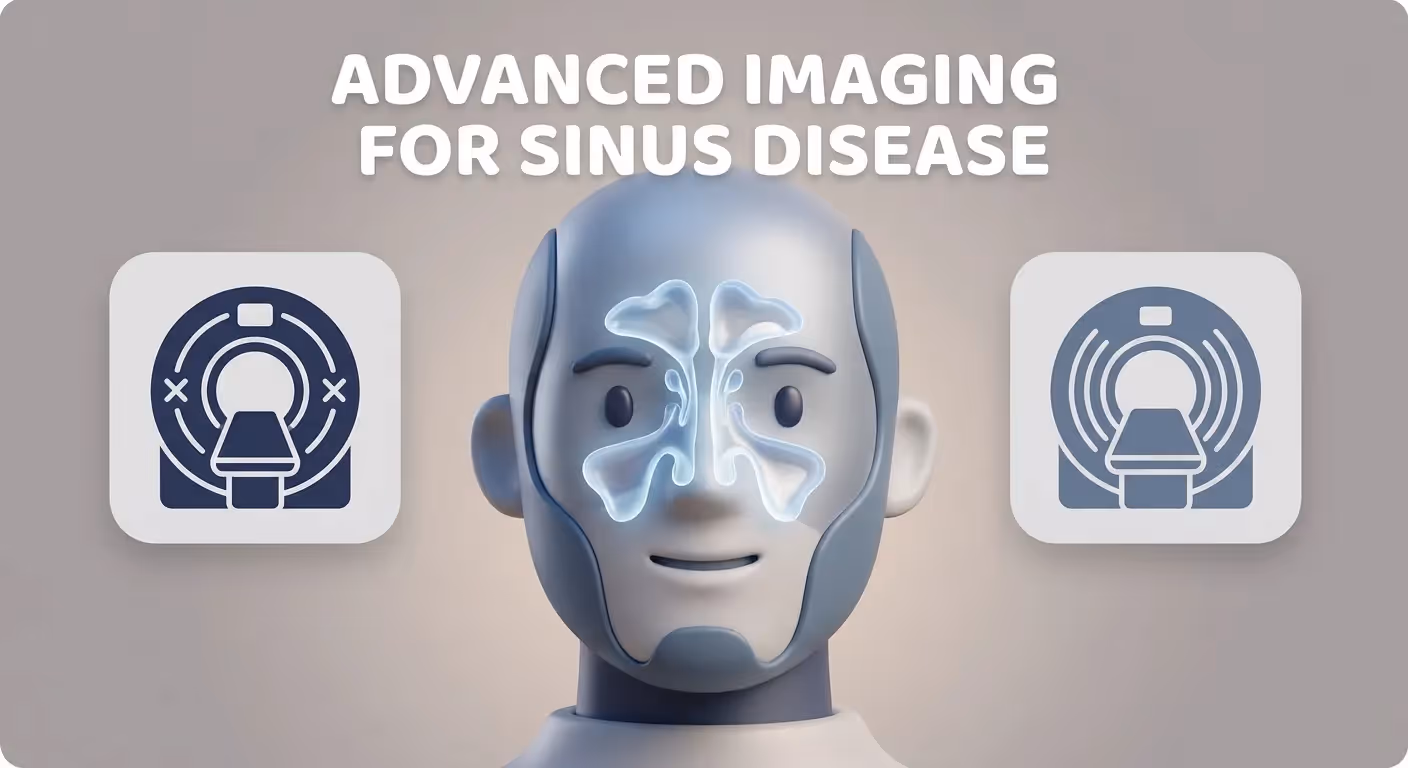
When Advanced Imaging Is Needed for Sinus Disease: CT and MRI Diagnosis Guide
If you’ve had sinus symptoms long enough, it’s natural to wonder whether a scan would “finally show what’s going on.” The truth is that advanced imaging for sinus disease (CT and MRI) is incredibly helpful in the right situations—and unnecessary (and sometimes misleading) in others.
A helpful way to think about imaging is this: a scan is a tool, not a diagnosis. The best scans answer a specific clinical question—not a general sense of “something feels off.”
Below is a clear, patient-friendly guide to when imaging is typically considered, what a sinus CT scan or sinus MRI can (and can’t) show, and how results are used to guide next steps.

Why most sinus problems don’t need imaging right away
Most acute, uncomplicated rhinosinusitis (the common “sinus infection” after a cold) is diagnosed based on your history and exam, not scans. In many routine cases, imaging doesn’t change treatment and can create confusion—because scans may show abnormalities even when symptoms are mild or improving, which can lead to unnecessary concern. This conservative approach is reflected in clinical guidance from the American Academy of Family Physicians (AAFP): imaging is not routinely recommended for uncomplicated acute sinusitis (AAFP, 2002: https://www.aafp.org/pubs/afp/issues/2002/1115/p1882.html).
A key principle: imaging is most meaningful when it matches the clinical picture (symptoms + exam). Reviews also emphasize avoiding “scan-first” decisions because CT findings are common even in people without significant sinus symptoms (Cleveland Clinic Journal of Medicine, 2020: https://www.ccjm.org/content/87/8/485).
Common reasons imaging is overused (and why doctors avoid it)
- Incidental findings are common. Mild mucosal thickening (swelling of the lining) can appear on CT in people who feel fine—or in someone who just had a cold and is already improving.
- Imaging can’t reliably distinguish viral vs bacterial infection in routine acute cases. That decision is usually clinical.
- A scan can “look scary” without being urgent. Seeing the word opacification on a report can be alarming, even when it simply reflects inflammation that often responds to routine care.
*Bottom line: If a scan won’t change the plan, it’s usually better to wait and focus on symptoms, exam, and response to treatment first.*
Sinus symptoms that should trigger a deeper evaluation
Use this as a checklist. Symptoms don’t automatically mean you need a scan, but they often guide whether further evaluation—including advanced imaging for sinus disease (CT and MRI)—might be discussed.
A quick “clinician-style” question that often determines next steps is: Are we dealing with a short-term flare… or a longer-term pattern that keeps coming back or never fully clears?
Symptoms that may suggest chronic or recurrent sinus disease
- Nasal congestion/obstruction lasting weeks
- Facial pressure/fullness
- Thick nasal drainage or postnasal drip
- Reduced smell/taste
- Symptoms that keep returning after seeming to improve (suggesting recurrent acute sinus infections)
Example: you feel better for a week or two, then the same cycle of pressure + drainage returns repeatedly—especially if you’re needing multiple rounds of medication to “get back to baseline.”
“Red flag” symptoms that can suggest complications (urgent evaluation)
- Swelling/redness around the eye, vision changes, severe headache
- High fever, neck stiffness, confusion, neurologic symptoms
- Severe facial swelling or rapidly worsening pain
These can signal complicated sinusitis, where clinicians often escalate evaluation quickly, including imaging when appropriate (Medscape overview: https://emedicine.medscape.com/article/384649-overview).
*Bottom line: Persistent, recurrent, or red-flag symptoms deserve prompt clinician evaluation—and sometimes targeted imaging.*

When advanced imaging is recommended (the big 4 scenarios)
Imaging is most often considered in these evidence-based situations. In plain language: when the answer will change what you do next.
1) Chronic rhinosinusitis (CRS): symptoms ≥ 12 weeks
Chronic rhinosinusitis generally means sinus-type symptoms most days for 3 months or more. Imaging may be used to:
- Confirm the extent and pattern of inflammation
- Identify anatomic blockage or drainage patterns
- Support longer-term treatment decisions, including procedural planning when relevant
(AAFP, 2002: https://www.aafp.org/pubs/afp/issues/2002/1115/p1882.html; CCJM, 2020: https://www.ccjm.org/content/87/8/485)
Learn more about chronic sinusitis here: https://sleepandsinuscenters.com/chronic-sinusitis
2) Recurrent acute sinus infections
A recurrent pattern often means repeated “acute” episodes with significant improvement (or full resolution) between them. A scan may help evaluate for:
- Structural narrowing that affects drainage
- Persistent disease between infections
Note: Imaging is complementary to clinical evaluation and not definitive on its own. (Medscape overview: https://emedicine.medscape.com/article/384649-overview)
3) Symptoms that persist despite appropriate treatment
When symptoms don’t improve as expected, imaging can help look for:
- Nasal polyps or chronic inflammation patterns
- Disease in less obvious areas (such as the sphenoid sinus)
- Ongoing obstruction that may change the treatment strategy
4) Suspected complicated infection (spread to the eyes or brain)
This is one of the most important reasons for urgent imaging. The goal is to quickly check for extension beyond the sinuses—especially toward the orbit (eye area) or intracranial space. Depending on the concern, clinicians may use CT and/or MRI (Medscape: https://emedicine.medscape.com/article/384649-overview).
*Bottom line: Imaging is most useful when it answers a specific question that will change your care plan.*

CT vs MRI for sinus disease—what’s the difference?
Here’s the simplest way to remember it:
- CT = “bony roadmap.”
- MRI = detailed soft-tissue imaging (mucosa, nerves, and potential masses), with superior evaluation of suspected orbital or intracranial involvement.
This division is widely reflected in radiology and clinical reviews (RadiologyInfo: https://www.radiologyinfo.org/en/info/sinusct; CCJM, 2020: https://www.ccjm.org/content/87/8/485).
Sinus CT scan
Best for: Bone, anatomy, and drainage pathways
Common uses: Pre-surgical sinus CT, FESS planning, evaluating chronic/recurrent patterns
Sinus MRI
Best for: Soft-tissue detail
Common uses: Tumor vs inflammation questions, orbital/brain involvement, atypical findings
For a deeper explanation of CT findings, see: sinus CT scan (what it shows and how it helps) https://sleepandsinuscenters.com/blog/sinus-ct-scan-what-it-shows-and-how-it-helps-diagnose-sinus-issues
*Bottom line: CT maps the pathways; MRI characterizes the tissues—your provider chooses based on the question at hand.*

What a sinus CT scan shows (and when it’s the best choice)
A sinus CT scan is often the go-to test when the main question is sinus anatomy and drainage.
A clinician might phrase the reason for CT like this: “Let’s map what your sinuses look like and where the bottleneck is.”
What CT is best at detecting
- Sinus opacification (areas filled with fluid or thickened lining)
- Narrow drainage pathways and anatomic contributors (septal deviation, turbinate variations, concha bullosa)
- Bony changes from long-standing inflammation
- Detailed anatomy mapping used for procedures
(RadiologyInfo: https://www.radiologyinfo.org/en/info/sinusct)
CT for pre-surgical planning (FESS and balloon sinuplasty)
For procedure planning, CT functions like a GPS map of the sinus pathways and the important nearby structures. This is why a pre-surgical sinus CT is commonly used for:
- FESS planning (functional endoscopic sinus surgery)
- Balloon sinuplasty CT evaluation and candidacy discussions
(CCJM, 2020: https://www.ccjm.org/content/87/8/485)
If you’re researching options, this overview may help: balloon sinuplasty https://sleepandsinuscenters.com/balloon-sinuplasty
CT limits (what it can’t tell you reliably)
- Whether symptoms are viral vs bacterial in routine acute illness
- Whether a CT finding is the cause of symptoms without clinical correlation
This is why clinicians emphasize matching CT results to symptoms and exam findings (CCJM, 2020: https://www.ccjm.org/content/87/8/485).
*Bottom line: CT is excellent for seeing structure and blockage, but it requires clinical context to guide action.*

What a sinus MRI shows (and when it’s the best choice)
A sinus MRI is typically used when the priority is soft-tissue characterization rather than fine bony anatomy.
One way to think of MRI: it helps clarify what something is made of (soft tissue detail), especially when the situation is atypical or potentially more serious.
When MRI is preferred
- Concern for spread beyond the sinuses (orbit/brain)
- Concern for tumor, invasive fungal disease, or atypical soft-tissue processes
- Situations where distinguishing retained secretions vs a soft-tissue mass is important
(CCJM, 2020: https://www.ccjm.org/content/87/8/485; Medscape: https://emedicine.medscape.com/article/384649-overview)
MRI limits (what it may not show as well as CT)
MRI usually does not display the small bony structures and drainage anatomy as clearly as CT—so CT is often preferred when surgery planning is the primary goal.
*Bottom line: MRI shines when soft-tissue detail and possible complications are the key questions.*
“Imaging alone isn’t enough”—how doctors interpret CT/MRI results correctly
Why scans can look abnormal even if you feel fine
Mild mucosal thickening can be common, especially after a recent cold or allergy flare. That’s why advanced imaging for sinus disease (CT and MRI) is interpreted alongside:
- Symptoms (pattern, duration, severity)
- Nasal exam findings
- Sometimes endoscopy findings
(Clinical correlation emphasis: CCJM, 2020: https://www.ccjm.org/content/87/8/485)
A practical analogy: a scan is like a photo of traffic. It tells you what the road looks like at that moment—but you still need the full story (timing, pattern, exam) to know why the traffic is there and what to do about it.
What else may be done with or instead of imaging
- Nasal endoscopy (an in-office look inside the nasal cavity)
- Allergy evaluation if triggers are suspected
- Medication trials and follow-up symptom scoring over time
*Bottom line: Scans are one piece of the puzzle—your symptoms and exam complete the picture.*
What to expect during a sinus CT or MRI (step-by-step)
Preparing for a sinus CT
- Usually very quick (often just minutes)
- You may be asked to remove metal objects
- Typically no special prep for routine sinus CT
(RadiologyInfo: https://www.radiologyinfo.org/en/info/sinusct)
Preparing for a sinus MRI
- Longer test time than CT and louder sounds
- Screening questions about implants/metal are important
- Some people discuss comfort options if claustrophobia is a concern
Contrast dye—when it’s used and what patients should know
- CT contrast is not always needed for routine sinus CT, but may be used for specific questions.
- MRI contrast may be used when evaluating complications, tumors, or atypical findings.
- Contrast agents are generally safe but carry small risks, including allergic reactions and kidney effects, so their use is carefully considered. Clinicians typically review kidney history and prior contrast reactions when contrast is being considered.
*Bottom line: Most sinus CTs are quick and non-contrast; contrast is added only when the benefits clearly outweigh the risks.*
Safety considerations: radiation, pregnancy, and other concerns
Is a sinus CT scan a lot of radiation?
CT uses ionizing radiation, and many modern sinus CT protocols aim to keep dose as low as practical while still getting usable images. A helpful decision-making question is: “Will this test change my care plan?” If the answer is yes, imaging is more likely to be worth it.
For more detail, see: radiation dose in sinus CT scans https://sleepandsinuscenters.com/blog/radiation-dose-in-sinus-ct-scans-what-you-need-to-know
Imaging considerations in pregnancy and children
In general, clinicians try to avoid unnecessary radiation when possible. Pregnant patients should always inform their healthcare provider before imaging to ensure the safest approach; when feasible, MRI or ultrasound may be preferred. Imaging choices for children are individualized to minimize exposure while answering the clinical question.
*Bottom line: Safety is built into modern protocols—share pregnancy status and other concerns with your care team so they can tailor the plan.*
Treatment decisions after imaging (what happens next)
Imaging doesn’t treat sinus disease—but it can clarify the diagnosis and guide a plan.
If CT/MRI supports chronic sinusitis
- Saline irrigation
- Topical nasal steroid sprays or rinses
- Addressing contributing factors (allergies, irritants)
- In selected cases, additional medications or procedure discussions
If imaging suggests an anatomic blockage
- Options often involve weighing continued medical therapy vs a procedure
- Balloon sinuplasty in appropriate candidates
- FESS when disease is extensive, persistent, or associated with significant obstruction/polyps
If imaging raises concern for complications
This may shift care toward urgent evaluation and coordinated specialty management depending on severity and findings (Medscape: https://emedicine.medscape.com/article/384649-overview).
*Bottom line: Imaging helps personalize your plan—medical therapy for many, procedures for the right candidates, and urgent care when complications are suspected.*
Lifestyle tips to support sinus health (with or without imaging)
Daily habits that reduce flare-ups
- Saline rinses (use safe water practices: sterile/distilled or previously boiled and cooled)
- Humidity control (avoid overly dry air)
- Smoke/fragrance avoidance when they trigger symptoms
Allergy-trigger control (when relevant)
For seasonal or perennial patterns, allergy evaluation and targeted treatment can be part of a longer-term strategy.
*Bottom line: Simple daily steps can reduce flares and support any treatment plan your clinician recommends.*
FAQs
Do I need a CT scan for a sinus infection?
Often no for acute, uncomplicated cases. Imaging is more commonly considered for chronic sinusitis diagnosis, recurrent acute patterns, suspected complications, or persistent symptoms that don’t improve as expected (AAFP, 2002: https://www.aafp.org/pubs/afp/issues/2002/1115/p1882.html).
What’s better for sinus problems—CT or MRI?
It depends on the question. CT is typically best for anatomy and surgery planning; MRI is typically best for soft-tissue concerns and suspected complications (CCJM, 2020: https://www.ccjm.org/content/87/8/485).
What if my CT scan is “normal” but I still have symptoms?
A normal scan can happen even with real symptoms, especially if the cause is not primarily sinus inflammation (for example: rhinitis, certain headache syndromes, reflux-related irritation, or other facial pain sources). This is one reason clinical correlation is emphasized (CCJM, 2020: https://www.ccjm.org/content/87/8/485).
Can a CT scan tell if it’s viral or bacterial?
Not reliably in uncomplicated acute disease. That distinction is usually based on symptom pattern and exam findings rather than imaging.
Will insurance cover a sinus CT or MRI?
Coverage is common when documentation supports medical necessity (chronic symptoms, recurrent acute infections, suspected complications, or pre-op planning). Prior authorization may apply depending on the plan.
When to see an ENT (call-to-action)
Consider an ENT evaluation at Sleep and Sinus Centers of Georgia if you have:
- Symptoms lasting 12 weeks or longer
- Multiple infections per year (recurrent pattern)
- Any red-flag symptoms (eye swelling, vision changes, severe headache, confusion/neurologic symptoms)
- Interest in procedure options where a pre-surgical sinus CT is part of planning (including FESS planning or balloon options)
Used appropriately, advanced imaging for sinus disease (CT and MRI) can be a powerful tool—not as a first step for every cold, but as a targeted way to clarify chronic, recurrent, or complicated sinus concerns.
To take the next step, you can book an appointment here: https://www.sleepandsinuscenters.com/
This article is for educational purposes only and is not medical advice. Please consult a qualified healthcare provider for diagnosis and treatment.
Don’t let allergies slow you down. Schedule a comprehensive ENT and allergy evaluation at Sleep and Sinus Centers of Georgia. We’re here to find your triggers and guide you toward lasting relief.